Research
Speckle Contrast Optical Spectroscopy
Example of dynamical speckle patterns generated from a rotating diffuser with increasing speed.
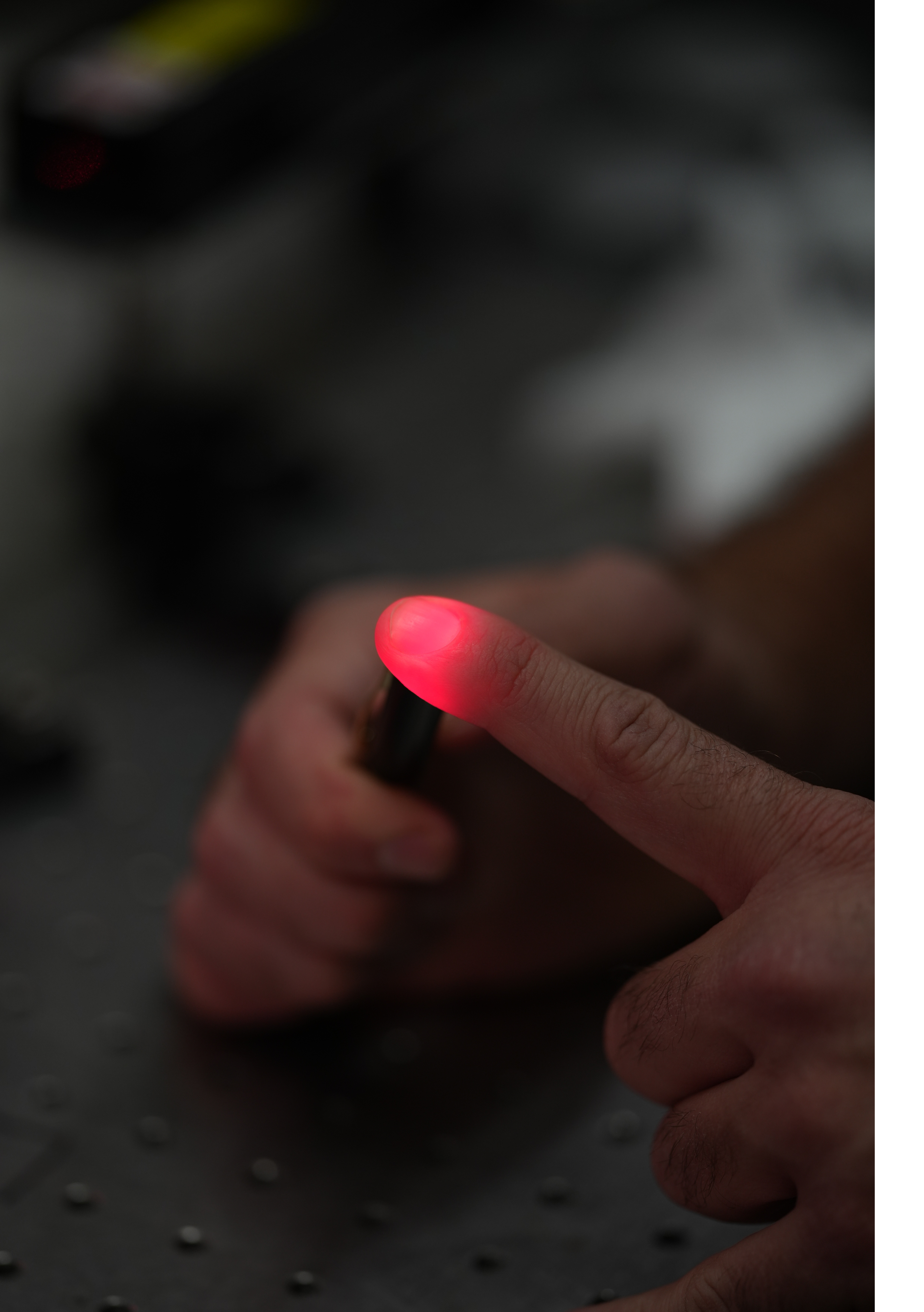
Speckle Contrast Optical Spectroscopy (SCOS) is a non-invasive optical technique that measures temporal fluctuations in speckle patterns formed by coherent light scattered from biological tissue. By analyzing changes in speckle contrast from sepckle patterns recorded over time by a camera typically working at 20 frames-per-second or more, SCOS provides information about blood flow dynamics and tissue perfusion at microvascular scales. In my research on SCOS, I developed and optimized systems to non-invasively measure blood flow dynamics in biological tissue such as the brain. By analyzing fluctuations in speckle patterns generated by coherent light interacting with moving scatterers, I was able to extract crucial physiological information, particularly related to cerebral hemodynamics. I focus on improving signal-to-noise ratios and minimizing motion artifacts, which are key challenges for accurate and robust SCOS measurements.
Blood flow and blood volume measuring speckle devices
I designed and tested advanced optical devices specifically tailored for measuring cerebral blood flow in both research and clinical environments. My work involved creating compact, wearable devices that utilize near-infrared light to penetrate the scalp and skull, allowing real-time monitoring of brain perfusion. I am particularly interested in suppressing superficial artifacts, ensuring that the signals we capture reflected true cortical blood flow changes, which is critical for applications like stroke monitoring or neurocritical care.
Chicken egg’s blood vessel imaging
Typical LSCI-recorded blood flow of a chick embryo.
As part of my exploration into biomedical imaging, I adapted optical techniques to visualize the vascular networks of developing chicken embryos, as shown in the video above. Using high-resolution laser speckle contrast imaging (LSCI), we were able to non-invasively monitor the dynamic blood flow through vessels as small as 100 µm in near real-time. We are now applying LSCI-based vascular imaging to a range of biological application including early sexing of chick embryos, predicting embryo survival rates, investigating the effects of antihypthensive drugs on vascular development, monitoring heart formation, and more.
Multi-modal degenerate laser cavity imaging
I investigated the use of a multi-modal degenerate laser cavity as a novel imaging platform capable of adapting to different imaging modalities. By controlling the spatial modes within the cavity, I demonstrated ways to enhance resolution, suppress noise, and even perform imaging through scattering media. This flexible laser system opened up new possibilities for highly efficient, tunable optical imaging, combining the benefits of traditional microscopy with the unique properties of complex laser cavities.

