
Research
Speckle Contrast Optical Spectroscopy
Example of dynamical speckle patterns generated from a rotating diffuser with increasing speed.
Speckle Contrast Optical Spectroscopy (SCOS) is a non-invasive optical technique that measures temporal fluctuations in speckle patterns formed by coherent light scattered from biological tissue. By analyzing changes in speckle contrast from sepckle patterns recorded over time by a camera, SCOS provides information about blood flow dynamics and tissue perfusion at microvascular scales. In my lab, we develop and optimize SCOS systems to non-invasively measure cerebral blood flow and volume in tissues such as the scalp, skull, or brain. Our long-term aim is to design a SCOS useful for medical application such as the prediction of having a stroke or the diagnosis of a brain injury.
Chicken egg’s blood vessel imaging
Typical LSCI-recorded blood flow of a chick embryo.
We adapted speckle imaging optical techniques to visualize developing chicken embryos, as shown in the video. Using laser speckle contrast imaging (LSCI), we were able to non-invasively monitor the dynamic blood flow through vessels as small as 70 µm in near real-time. Using this system, we are investigating a range of biological applications including cardiac angiogenesis, embryo development, non-invasive sexing, and drugs testing on vascular development.
Blood flow and blood volume measuring speckle devices
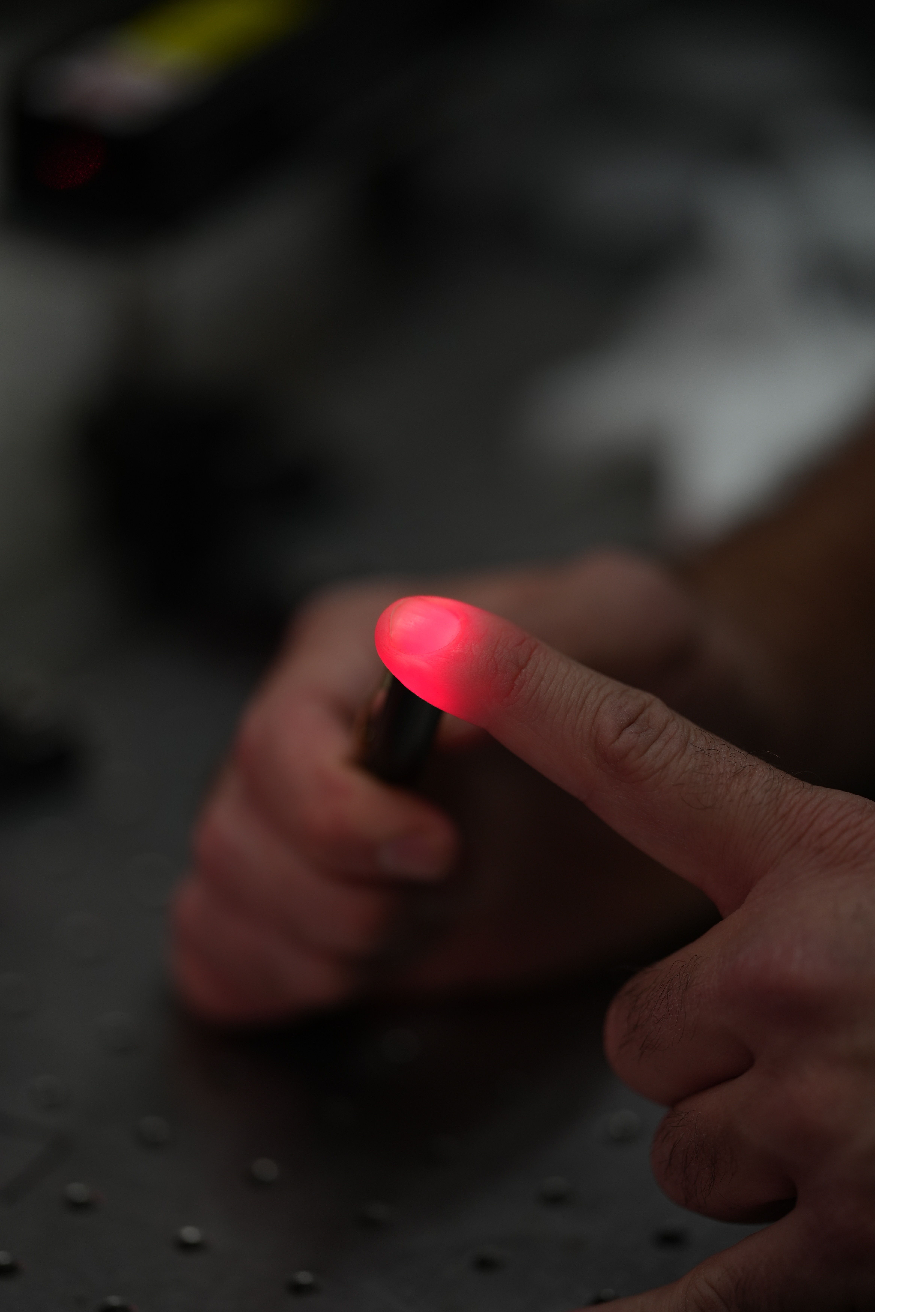
The lab also uses speckle imaging techniques (SCOS) to monitor blood flow and blood volume in different parts of the body. We are interested in exploring the potential of speckle-based imaging as a noninvasive tool for screening, monitoring, and potentially diagnosing a variety of diseases and medical conditions from different areas of the human body.
Multi-modal degenerate laser cavity imaging
The lab also has interest in designing multi-modal imaging laser sources. For example, a degenerate laser cavity can serve as a powerful imaging platform, with superior adaptability to different scenarios. By controlling the spatial modes within the cavity, one can enhance resolution, suppress noise, and perform imaging through scattering media. This flexible laser system open possibilities for highly efficient, tunable optical imaging, combining the benefits of traditional microscopy with the unique properties of complex laser cavities.